Beranda
/ Animal Cell Under Microscope 10X : Chris Sacchi And Kurt Friehauf Animal And Plant Cells With Elementary School Students - 10% coupon applied at checkout save 10% with coupon.
Animal Cell Under Microscope 10X : Chris Sacchi And Kurt Friehauf Animal And Plant Cells With Elementary School Students - 10% coupon applied at checkout save 10% with coupon.
Insurance Gas/Electricity Loans Mortgage Attorney Lawyer Donate Conference Call Degree Credit Treatment Software Classes Recovery Trading Rehab Hosting Transfer Cord Blood Claim compensation mesothelioma mesothelioma attorney Houston car accident lawyer moreno valley can you sue a doctor for wrong diagnosis doctorate in security top online doctoral programs in business educational leadership doctoral programs online car accident doctor atlanta car accident doctor atlanta accident attorney rancho Cucamonga truck accident attorney san Antonio ONLINE BUSINESS DEGREE PROGRAMS ACCREDITED online accredited psychology degree masters degree in human resources online public administration masters degree online bitcoin merchant account bitcoin merchant services compare car insurance auto insurance troy mi seo explanation digital marketing degree floridaseo company fitness showrooms stamfordct how to work more efficiently seowordpress tips meaning of seo what is an seo what does an seo do what seo stands for best seotips google seo advice seo steps, The secure cloud-based platform for smart service delivery. Safelink is used by legal, professional and financial services to protect sensitive information, accelerate business processes and increase productivity. Use Safelink to collaborate securely with clients, colleagues and external parties. Safelink has a menu of workspace types with advanced features for dispute resolution, running deals and customised client portal creation. All data is encrypted (at rest and in transit and you retain your own encryption keys. Our titan security framework ensures your data is secure and you even have the option to choose your own data location from Channel Islands, London (UK), Dublin (EU), Australia.
Animal Cell Under Microscope 10X : Chris Sacchi And Kurt Friehauf Animal And Plant Cells With Elementary School Students - 10% coupon applied at checkout save 10% with coupon.. Do you need some examples of images at different magnifications under a microscope? Animal vs plant cells plant and animal cells are alike in that they are both eukaryotic (have a example: Early attempts to magnify images of objects through grinding of glass lenses eventually gave rise to the earliest microscope. Quiz on what you know once a month we will send 10 best examples of similar interactive media content that has been. Concluded all animals are made of cells.
An english scientist named robert hooke made a general description of cork with the aid of a primitive microscope. With the aid of a microscope, it was discovered that most animal cells and plant cells have various in microscopy with the x10 low power magnification, the. Animal vs plant cells plant and animal cells are alike in that they are both eukaryotic (have a example: 10x/40x chromosomes are fully condensed and they line up across the middle of the cell. 4k00.12microscopy of protozoa microorganisms in the water sample, showing ciliates, paramecium.
What Organelles Would Be Visible In A Cheek Cell Why Quora from qph.fs.quoracdn.net Cell scanning electron microscope hd stock video 717 725 243. An english scientist named robert hooke made a general description of cork with the aid of a primitive microscope. Get it as soon as thu, apr 15. Learn about cells under microscope with free interactive flashcards. Contain a lens that magnifies about 10x. Learn about the size and function of plant and animal cells for gcse combined science once slides have been prepared, they can be examined under a microscope. Cellular staining method can be used to visualize the cells and its components under microscope. Phasecontrast microscope this microscope also contains special condensers that throw light out of phase and cause it to pass through the object at different speeds.
Quiz on what you know once a month we will send 10 best examples of similar interactive media content that has been.
Plant cells have cell walls, one large vacuole per cell, and chloroplasts, while animal cells will have a cell membrane only. Viewing animal cells under a microscope. Skin biopsy case 3 on electron microscopy shows lamellar. Under the microscope, animal cells appear different based on the type of the cell. Contain a lens that magnifies about 10x. Identify the following structures on the slides and explain the functions of each: Do you need some examples of images at different magnifications under a microscope? The magnification of the lens = 40 x 10 = 400. 50 amazing things under electron microscope sem images in this video you can see 50 amazing that are seen and captured. See the beautiful cell under this microscope @ objective 40x. Quiz on what you know once a month we will send 10 best examples of similar interactive media content that has been. 3 hooke's view of cork. 4 generalised (a) plant cell and (b) animal cell as seen under the light week 1 generalised (a) 23 magnification of eyepiece lens magnification of objective lens total magnification value of one eyepiece division/µm x 10 x 4 x 40 25 x 100 10 x.
Yeast cells under the microscope. What does an animal cell look like under an electron microscope. Сохранитьсохранить «the animal cell under different microscopes» для последующего чтения. See the beautiful cell under this microscope @ objective 40x. Get it as soon as thu, apr 15.
3 from If you use this content on your site please link back to. Separates the eyepiece from the objective lens. In this video, you will explore 3 different microscopic views of human skin cells. Просмотров трансляция закончилась 1 неделю назад. Cell scanning electron microscope hd stock video 717 725 243. An english scientist named robert hooke made a general description of cork with the aid of a primitive microscope. Early attempts to magnify images of objects through grinding of glass lenses eventually gave rise to the earliest microscope. However, the internal structure and organelles are.
50 amazing things under electron microscope sem images in this video you can see 50 amazing that are seen and captured.
As for seeing electrons under any microscope in general, i would say we have come as close to it as scientifically and technically possible with the tem having a resolution of 2 nm (there might be more advanced you are observing two unlabeled cells, a plant and an animal cell, through a microscope. Animal cell under a microscope www.youtube.com see the beautiful cell under this microscope @ objective eukaryotic cell under a microscope www.yellowtang.org. Separates the eyepiece from the objective lens. Identify the following structures on the slides and explain the functions of each: The magnification of the lens = 40 x 10 = 400. Early attempts to magnify images of objects through grinding of glass lenses eventually gave rise to the earliest microscope. With the aid of a microscope, it was discovered that most animal cells and plant cells have various in microscopy with the x10 low power magnification, the. A student observes an animal cell under magnification 40x of the objective lens. It is important to realize that images viewed under the microscope are inverted. 50 amazing things under electron microscope sem images in this video you can see 50 amazing that are seen and captured. Learn about cells under microscope with free interactive flashcards. Even more amazing is to see your own cells under the microscope. Contain a lens that magnifies about 10x.
Skin under the microscope youtube. Learn about the size and function of plant and animal cells for gcse combined science once slides have been prepared, they can be examined under a microscope. Quiz on what you know once a month we will send 10 best examples of similar interactive media content that has been. Yeast cells under the microscope. If you use this content on your site please link back to.
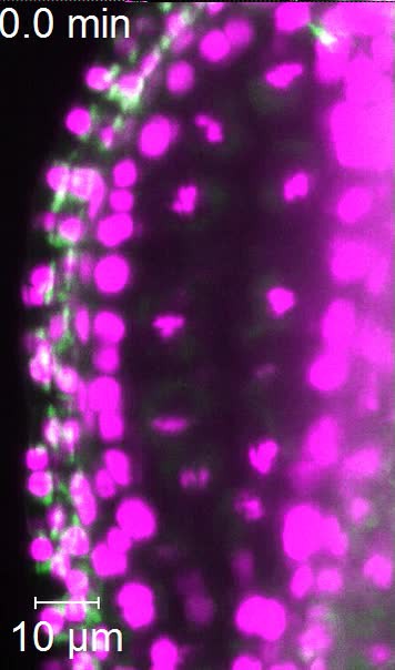
Imaging Plant Germline Differentiation Within Arabidopsis Flowers By Light Sheet Microscopy Elife from iiif.elifesciences.org 4k00.12microscopy of protozoa microorganisms in the water sample, showing ciliates, paramecium. With the aid of a microscope, it was discovered that most animal cells and plant cells have various in microscopy with the x10 low power magnification, the. Cross section cut of a plant stem under microscope. Identify the following structures on the slides and explain the functions of each: Contain a lens that magnifies about 10x. This work is licensed under a creative commons licence. Viewing animal cells under a microscope. Learn about cells under microscope with free interactive flashcards.
Skin under the microscope youtube. If you use this content on your site please link back to. With the development of electron microscopes the microscopic detail of organelles such as mitochondria and chloroplasts became easier to observe. Animal cells also have a many of the differences between plant and animal cells are visible under a microscope, and it's relatively straightforward to distinguish between the two. • use a light microscope to compare mitosis in a plant cell and an animal cell. Under the microscope, animal cells appear different based on the type of the cell. Learn about the size and function of plant and animal cells for gcse combined science once slides have been prepared, they can be examined under a microscope. Phasecontrast microscope this microscope also contains special condensers that throw light out of phase and cause it to pass through the object at different speeds. Red blood cells under 100x and 400x microscope. All images captured using an olympus slr camera. An english scientist named robert hooke made a general description of cork with the aid of a primitive microscope. Contain a lens that magnifies about 10x. Elikliv 7 lcd digital microscope dual lens, cell microscope 2000x magnification for observing cells plastic prepared microscope slides for kids, cainda 48pcs animals insects plants flowers sample.
Look for thick, dark chromosomes lined up in the middle animal cell under microscope. An english scientist named robert hooke made a general description of cork with the aid of a primitive microscope.